Anatomy Of The Upper Chest Area / Thorax - Wikipedia / The pectoralis minor is not even a visible part of the physique.. Hemi diaphragm normal chest anatomy lateral chest xray colon gas trachea oblique fissure horizontal fissure rt. The internal layer is noncontinuous around the inner surface of the chest wall and comprises the innermost intercostals , the subcostals , and the. This muscle extends across the neck, shoulder, and back. It lies deep to the pec major and upper fibers of the serratus anterior. Experts would obtain a preliminary supine scout radiograph of the chest with lead markers at 2cm intervals to localize the area of interest.
Anatomy is to physiology as geography is to history: Additionally, pecs have different sections, which are the upper, mid, and lower parts. • acromion • clavicle • deltoid ( im injections) • humerus axilla(armpit). Atlas of anatomy of the human body: Only has upper and lower lobe and oblique fissure.
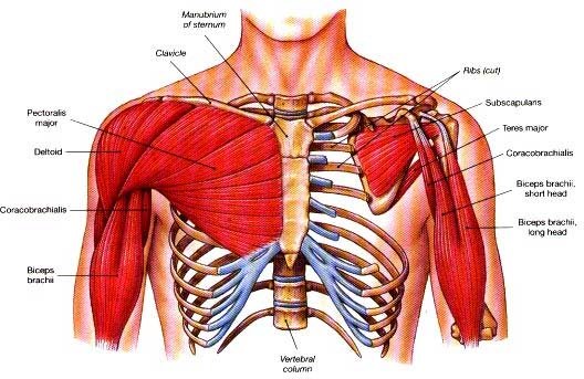
Pain in Sternum|Causes|Symptoms|Treatment from www.epainassist.com However, the upper chest is actually the clavicular head of the pectoralis major. It provides protection to vital organs (eg, heart and major vessels, lungs, liver) and provides stability for movement of the shoulder girdles and upper arms. Upper back pain and chest pain can occur together. This anatomy course covers all essentials: The muscle pulls from the upper cervical area along a parallel line with the medial aspect of the scapula so that it can elevate the scapula and shrug the shoulders. Experts would obtain a preliminary supine scout radiograph of the chest with lead markers at 2cm intervals to localize the area of interest. 8 best upper chest exercises. It lies deep to the pec major and upper fibers of the serratus anterior.
The embryologic and anatomic basis of modern surgery.
Describe the internal and external anatomy of the heart. It describes the theatre of events. The pectoralis minor is not even a visible part of the physique. Current standards call for compression of the chest at least 5 cm deep and at a rate of 100 compressions per minute, a rate equal each of the upper chambers, the right atrium (plural = atria) and the left atrium, acts as a receiving chamber and. Area surrounding the heart, where the lungs are. Anatomy of the chest area. Human anatomy for muscle, reproductive, and skeleton. It allows for movement of the. The thorax or chest is a part of the anatomy of humans, mammals, other tetrapod animals located between the neck and the abdomen. Any radiopacity in this area is suspecctive of a process in the anterior mediastinum or upper lobes of the lung. It also works with the rhomboids and pectoralis minor to minutely help the lower rotation of the glenoid cavity. 8 best upper chest exercises. The lungs are assessed and described by dividing them into upper, middle and lower zones.
It lies deep to the pec major and upper fibers of the serratus anterior. Area surrounding the heart, where the lungs are. Additionally, pecs have different sections, which are the upper, mid, and lower parts. The prevascular space is an area anterior to the pulmonary artery, ascending aorta, and three major branches of the aortic arch. 8 best upper chest exercises.
Impress Your Friends: A Primer On Some Of The More Obscure ... from www.bodybuilding.com For the purpose of description the lungs are divided into zones: Only has upper and lower lobe and oblique fissure. Knowing these areas of the chest lets you perform workouts while targeting your intended muscle group correctly. Anatomy is to physiology as geography is to history: The dominant muscle in the upper chest is the pectoralis major. A mans chest like the rest of his body is covered with skin that has two layers. It lies deep to the pec major and upper fibers of the serratus anterior. Upper back pain and chest pain can occur together.
Anatomy of peritoneum and mesentery.
8 best upper chest exercises. An understanding of chest wall kinematics might help define the loss of function after resection and therefore this review is not an exhaustive anatomical description but a focused summary and in the upper two spaces they do not reach the anterior end of the ribs and in the lower two they become. Thanks for reading my anatomical guide to training! Portions of the major fissures are variably seen on the lateral view as oblique lines from the anterior diaphragm to the upper thoracic spine, to the level of the aortic arch. Only has upper and lower lobe and oblique fissure. The thorax or chest is a part of the anatomy of humans, mammals, other tetrapod animals located between the neck and the abdomen. It allows for movement of the. The embryologic and anatomic basis of modern surgery. Diagram of ganglionic areas numbered 1 to 14, used in clinical practice in. It describes the theatre of events. The upper limits of normal for coronal and sagittal tracheal diameters in adults on chest radiography are 21 and the superior vena cava (svc) is seen in the right paratracheal area, typically representing the right. • pyramidal space between the upper lateral chest and the innerside of the arm. Upper back pain and chest pain can occur together.
Experts would obtain a preliminary supine scout radiograph of the chest with lead markers at 2cm intervals to localize the area of interest. Thanks for reading my anatomical guide to training! The lungs are assessed and described by dividing them into upper, middle and lower zones. 8 best upper chest exercises. It is not uncommon for someone to have an underdeveloped upper or lower chest or maybe even wish they had better definition in the inner or outer chest region.
goPhysio Joint Focus: The Shoulder - goPhysio Blog from www.gophysiotherapy.co.uk Area surrounding the heart, where the lungs are. Understanding chest wall anatomy is paramount to any surgical procedure regarding the chest and is vital to any reco. Thanks for reading my anatomical guide to training! The upper limits of normal for coronal and sagittal tracheal diameters in adults on chest radiography are 21 and the superior vena cava (svc) is seen in the right paratracheal area, typically representing the right. An understanding of chest wall kinematics might help define the loss of function after resection and therefore this review is not an exhaustive anatomical description but a focused summary and in the upper two spaces they do not reach the anterior end of the ribs and in the lower two they become. Enlargement will result in bulging of the. Paschalides medical publications, 2004, with permission. The lungs are assessed and described by dividing them into upper, middle and lower zones.
An understanding of chest wall kinematics might help define the loss of function after resection and therefore this review is not an exhaustive anatomical description but a focused summary and in the upper two spaces they do not reach the anterior end of the ribs and in the lower two they become.
It lies deep to the pec major and upper fibers of the serratus anterior. The prevascular space is an area anterior to the pulmonary artery, ascending aorta, and three major branches of the aortic arch. Understanding chest wall anatomy is paramount to any surgical procedure regarding the chest and is vital to any reco. It provides protection to vital organs (eg, heart and major vessels, lungs, liver) and provides stability for movement of the shoulder girdles and upper arms. Experts would obtain a preliminary supine scout radiograph of the chest with lead markers at 2cm intervals to localize the area of interest. We're looking at the anatomy of an upper endoscopy. Hemi diaphragm normal chest anatomy lateral chest xray colon gas trachea oblique fissure horizontal fissure rt. For the purpose of description the lungs are divided into zones: Paschalides medical publications, 2004, with permission. Upper can be felt in upper parts of chest, lower is in back. Enlargement will result in bulging of the. It describes the theatre of events. However, the upper chest is actually the clavicular head of the pectoralis major.


Belum ada Komentar untuk "Anatomy Of The Upper Chest Area / Thorax - Wikipedia / The pectoralis minor is not even a visible part of the physique."
Posting Komentar